Monday, January 31, 2011

Fistuliporoid Bryozoans from the Silica Shale

I'm sure I've mentioned before about how difficult it can be to ID some Bryozoans beyond the Family level. Such is the case with some Fistuliporoid Bryozoans that I've found in the Silica shale.

Some form finger like branches


Others, massive colonies


Still others colonize Brachiopod shells


Without thin sections both perpendicular and parallel to the grain of the colony, I'll never be able to truly ID these even to a genus level. So they remain within my collection simply labelled "Fistuliporoid Bryozoan".

Saturday, January 29, 2011

Megastrophia concava from the Silica Shale

One of the largest Brachiopods found within the Silica Shale is Megastrophia concava. Superficially it looks like an overgrown Strophodonta, but the size is the key. No other Strophomenids grow this large within the Silica Shale and the thin radiating ribs on the surface are spaced farther apart than on any Strophodonta.


This is an partial brachial valve that shows the two kidney bean shaped muscle attachement scars.


This is a very easy shell to ID as Megastrophia concava based on the 1975 book by Robert V. Kesling & Ruth B. Chilman: Strata and Megafossils of the Middle Devonian Silica Formation: Museum of Paleontology, Papers on Paleontology, No. 8, pg. 96, pl. 26.

Thursday, January 27, 2011

Internal shell details of Strophodonta

While going through my finds from the Silica Shale of Sylvania, OH I came across a number of individual valves from Strophodonta. They are preserved well enough to show the internal structure of the shells where they muscles were attached.

This is a Brachial valve with some large muscle attachment structures. At the very top of the shell is a "Y" shaped structure that fit into a corresponding notch in the Pedicle valve. The main muscles that opened and closed the shell were anchored to the central scars.


This is the interior of a Pedicle valve and you can see some palmate scars in the shell structure that correspond to the raised areas of the Brachial valve pictures above. Notes the two kidney shaped, shallow depressions near the middle of the shell. This is where the "Y" shaped structure from the Brachial valve would be when the shells were together.


This is a different shell than the one pictured above. I chose this shell since it shows more of the hinge and the teeth that were used to help connect the Pedicle and Brachial valves together.

Tuesday, January 25, 2011

Strophodonta from Sylvania

I've featured Strophodonta shells from Sylvania before on my blog, in a post about the first fossils I bought, but I thought it would be useful to do a "turn-around" to show what it looks like from different angles.

Here are three specimens that show the variation in size and shape.


This is the largest specimen on the left side of the above picture. Note that it has a very deep concave shape. This is not limited to shells this size and as such could be a variation in growth or perhaps a species or subspecies.


A smaller specimen that is on the lower right of the first picture. Less concave that the previous specimen but greater hinge width when compared to shell length.


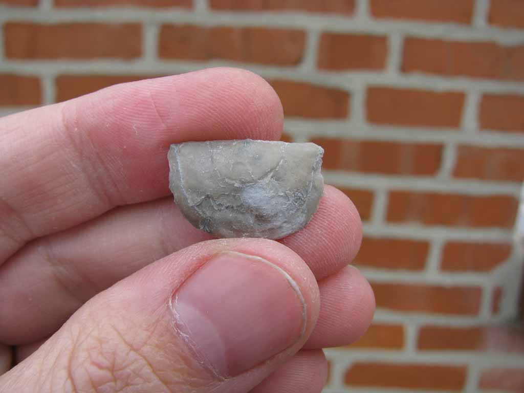
I picked this specimen off the piles specifically because the Pyrite replacement of it's shell had not completely oxidized and also because it afforded me a cut away view inside the shell. Most of the Strophodonta shells I collected loose are whole or partially crushed so that you can't get a good idea of the internal space within the shell.


Many Strophomenids, like Strophodonta, did not have hard parts to support it's Lophopores (feeding organs) like Atrypids or Spiriferids. As such there was no need to build a shell to store them.

This is pure conjecture but judging from the narrow space that can be seen in the above side view, the animal inflated it's body and organs with water when it opened it's shell. When they sensed a threat or otherwise closed, they would eject the water and their soft bodies would once again fit within the narrow confines of the shell. Or perhaps they pushed their lophopores out into the current to catch food particles.

Here are two separate valves (non associated) that I've arranged to illustrate what the animal would have looked like when opening it's shell to feed.

Looking at the exterior of the Brachial valve so you can see how the valves may have once fit together (were they from the same animal).


Side view


Front view


These specimens were collected from the Silica Shale dumps in Sylvania, Ohio in 2004.

Sunday, January 23, 2011

Random hash plates from the Silica Shale

The megafauna of the Silica shale gets all the attention when I'm looking for fossils in the field. When I'm at a site I've been consciously trying to pick up pieces of the bedrock to bring back a study. One reason is to better understand the paleo-environment that the animals lived in and another is to search for smaller members of the community. These plates are often referred to as "Hash" plates in an effort to describe the large quantity of shelly debris that is often present. Presented for your viewing pleasure are three small matrix pieces. While the pictures are not of the best quality, see if you can pick out the following fossils:

Tentaculites
Hyolites
Phacops rana
Chonetes
Ostracods
Crinoid stem segments
Sulcoretipora
Bryozoans


Friday, January 21, 2011

Protoleptostrophia from Sylvania

As I was going through boxes of Brachiopod shells from the Silica Shale I came across these thin, delicate fossils of Protoleptostrophia. I often find these Brachiopods in matrix and rarely loose like the below specimens. This is likely due to the extremely thin and delicate nature of the shells.

Pedicle Valve


Brachial Valve


These shells are so skinny I can't really get a good shot of the front, rear or sides like I do with other Brachiopods.

This was a loose shell of just the Pedicle Valve, but it shows the internal muscle scars.

Exterior


Interior


I'm going to call these Protoleptostrophia perplana based on the 1975 book by Robert V. Kesling & Ruth B. Chilman: Strata and Megafossils of the Middle Devonian Silica Formation: Museum of Paleontology, Papers on Paleontology, No. 8, pg. 99, pl. 13,24.

Wednesday, January 19, 2011

Pholidostrophia from Sylvania, OH

Pholidostrophia is a brachiopod that I was introduced to in the rocks along the Lake Erie shore at Eighteen Mile Creek. You can read those post by clicking here and another post of specimens from my local Mahantango formation here. Below are two specimens from the Silica Shale of Sylvania, Ohio that I collected a few years ago. It's interesting to compare the specimens from the similarly aged Mahantango formation and Wanakah Shale to these. The Silica Shale specimens are larger than those from the other two localities. This same feature seems to occur in many of the Brachiopod genera that are found in the Silica Shale and could be due to a calmer, more food rich environment in which they lived.

I'm going to call these Pholidostrophia geniculata based on the 1975 book by Robert V. Kesling & Ruth B. Chilman: Strata and Megafossils of the Middle Devonian Silica Formation: Museum of Paleontology, Papers on Paleontology, No. 8, pg. 100, pl. 118.

Pedicle Valve


Front


Brachial Valve


Rear


Profile


Another specimen

Pedicle Valve


Front


Brachial Valve


Rear


Profile

Monday, January 17, 2011

Sulcoretipora from the Silica Shale

Many Bryozoans are hard to ID due to their similar growth patterns and exterior look. Not so with the genera Sulcoretipora. As you can see in the pictures below it's a flat, branching Bryozoan. The only other common genera that has a similar look is Taeniopora but that has a prominent central rib that runs up the center of all the branches.


The specimen above all come from the Silica shale and were collected at Sylvania, OH.

I'm going to call these Sulcoretipora deissi based on the 1975 book by Robert V. Kesling & Ruth B. Chilman: Strata and Megafossils of the Middle Devonian Silica Formation: Museum of Paleontology, Papers on Paleontology, No. 8, pg. 71, pl. 44.

Saturday, January 15, 2011

Platycerid snails from the Silica Shale

Few snails are as well known in the Devonian as Platyceras is. The name sounds similar to that of an Ammonite since they share a similarly pronounced last part of their names. While they are both Mollusks, Platyceras is a Gastropod (or stomach foot) wile Ammonites are Cephalopods (head foot).

Platyceras is a very prolific genera within the Silurian to Devonian and is known to exist from the Late Ordovician (Sandbian - 460 mya) through the Permian (Capitanian - 260 mya). Within the Silica Shale they can grow to be quite large and I've found a couple of species:

Platyceras bucculentum (Hall 1862) - Note the undulating, wide, bell like margin. This species often has a well defined crest but this specimen is lacking that.


Another Platyceras bucculentum


This specimen appears to be a Platyceras rarispinum based on the spine bases present on the shell.


All ID's are based on the 1975 book by Robert V. Kesling & Ruth B. Chilman: Strata and Megafossils of the Middle Devonian Silica Formation: Museum of Paleontology, Papers on Paleontology, No. 8, pg. 117, pl. 19,20.